
Creating A Google Earth View of Cancer
A current focus in cancer is to develop new methods for monitoring multiple species in tissue, for example imaging drugs and cancer biomarkers under high-spatial resolution, to advance understanding of tumour development.
NPL is leading a team in developing the ‘Google Earth’ of tumour mapping – creating a reproducible, standardised way to fully understand different tumours in unprecedented detail
The ground-breaking project has been selected by Cancer Research UK to receive £16 million over the next five years – one of the biggest funding grants ever awarded by the charity – as part of its Grand Challenge awards, set up to revolutionise the prevention, diagnosis and treatment of cancer and help scientists attack some of the hardest, unanswered questions in cancer research.
Increasing biomedical imaging power with sub-cellular resolution. NPL is home to the UK’s National Centre of Excellence in Mass Spectrometry Imaging (NiCE-MSI). This premier centre provides powerful ‘eyes’ on the molecular and atomic world with beyond state-of-the-art imaging capability and plays a crucial role in improving repeatability and reproducibility of measurements.
Reducing the rate of drug failure is a major challenge for the pharmaceutical industry. The cost of developing a new medicine is approximately $1.8 billion2 because most candidate drugs fail to make it all the way through the development process. Those that fail at the late stage of drug development contribute the largest cost burden, so measurements at the early stages that can identify future failure are of great importance.
The measurement challenge is to see where drugs go at the sub-cellular level, to answer long-standing questions about whether drug concentrations are sufficiently high in the right places to have a therapeutic effect, or if the medicine is lodging within cellular components and causing toxicity. If anomalies are spotted earlier, it might help to explain toxicities or lack of efficacy of a medicine alongside reducing costly late-stage failures.
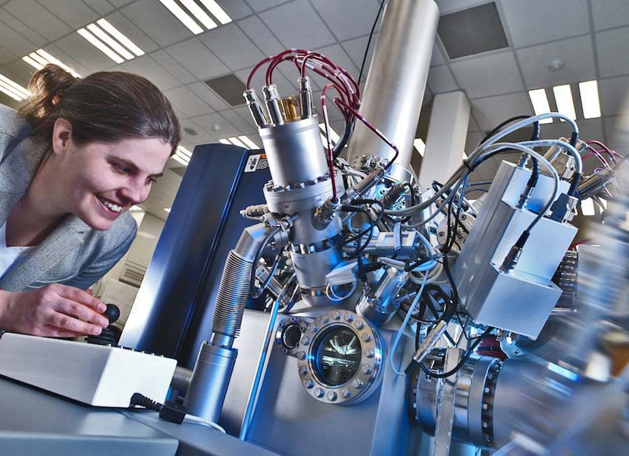
The NPL 3D OrbiSIMS, currently the only one of its kind in the world, is a revolutionary new instrument for 3D imaging of drug molecules and metabolites (such as lipids) with sub-cellular spatial resolution. It has the highest simultaneous spatial resolution and mass resolution (resolving power) worldwide. The instrument, unique to NPL, was developed in partnership with GlaxoSmithKline alongside leading instrument manufacturers. It will provide high-resolution biomedical imaging in the Cancer Research UK Grand Challenge project.
Accelerating nuclear medicine with CERN . Over a million patients undergo a nuclear medicine procedure in the UK each year, the majority of these administrations are for the diagnosis or staging of disease, including cancer3. The prominence of radionuclides is growing rapidly due to their ability to yield images of physiological function – an indispensable tool in cancer diagnosis. Radionuclides can be attached to carrier molecules that seek out cancerous cells thus allowing the tumour to be specifically destroyed with minimal effects on surrounding tissues.
NPL is working with CERN, one of the world’s largest and most respected research facilities, to develop novel radioisotopes for diagnostic, therapeutic and theranostic applications.
Delivering a better patient experience and increased accuracy in breast cancer diagnosis. Breast cancer is the most common cancer in the UK with 62,000 women being diagnosed each year. Current diagnosis relies on uncomfortable X-ray mammography, followed up by biopsy. Of all the lesions investigated, around 30% result in a malignant diagnosis thus some 70% of investigations are unnecessary. These invasive biopsies are estimated to cost the NHS £35 million annually. Conventional mammograms also rely on a radiologist or doctor’s interpretation and are less likely to detect breast cancer in younger or Asian populations, whom have denser breast tissue.
NPL is developing an ultrasound screening platform, currently entering clinical demonstration phase. This doesn’t use X-rays (so the patient isn’t exposed to radiation) and the screening is carried out with the breast submerged in warm water, without compression, which is a more comfortable experience for the patient. Its capability to better differentiate tissue properties should then ensure more accurate diagnosis.
Enabling greater confidence in radiotherapy for NHS England. Radiotherapy contributes to the cure of 40% of cancer patients cured of their disease. Most patients are treated using a high-energy beam of X-rays delivered to a precise area, using a linear accelerator (linac). All UK NHS external beam radiotherapy treatment doses are traceable to primary standards via NPL’s dosimetry calibration services.
The Metrology for Medical Physics Centre (MEMPHYS) at NPL partners with leading universities, hospitals and institutions including the Christie, the Royal Marsden, UCLH, Queen Elizabeth Hospital Birmingham and the Royal Surrey County Hospital, bringing together academics, manufacturers, scientists and clinicians to optimise cancer treatments. This optimisation helps to accelerate implementation of the most advanced techniques helping to ensure patients receive the same standard of treatment irrespective of where they receive their treatment.